
Description
Tissue samples, usually extracted from biopsies, surgeries, or excisions, are used for clinical diagnosis and prognosis for millions of patients each year. In 2014, Medicare, which covers approximately 30% of the United States population, reimbursed microscopic analysis of pathology samples nearly 20 million times, amounting to $964 million.
Our nuclei segmentation algorithm consists of three input algorithms: foreground marker detection, background marker detection, and gradient computation. As the latter is fairly straightforward, we need to investigate the effectiveness of our foreground and background marker algorithms. Figure 1 illustrates this, showing both the foreground markers (black regions in the center of each cell), and the background markers (the black lines).
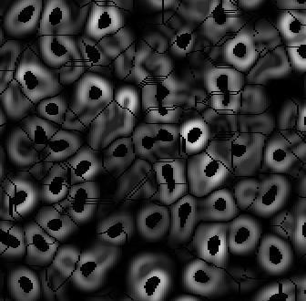
The gradient image, with the foreground markers and background markers (ridge lines) superimposed.
In this work, we have investigated the use of a supervised linear classifier to predict whether a pixel belongs to a nucleus based on its color. This is useful for a number of reasons.
1. It allows flexibility when dealing with different tissue types and staining protocols. For example, if a user is imaging breast or head and neck tissue, the same classifier would be able to adapt to changes in illumination, color schemes, and digital pathology scanners.
2. Through additional user feedback (e.g. the user identifies false positives and false negatives in the final segmentation), it is possible to refine the entire segmentation algorithm.

input image
MATLAB Implementation of segmentation-based disparity averaging
Reviews
There are no reviews yet.