Magnetic Resonance Imaging
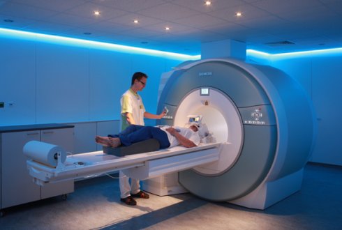
Magnetic Resonance Imaging (MRI) is a non-invasive and powerful imaging modality, with a broad range of applications in both clinical diagnosis and basic scientific research. Comparing to other medical imaging modalities, MRI does not use ionizing radiation and provides superior soft-tissue characterization with high resolution and flexible image contrast parameters. Moreover, MRI allows good visualization of anatomical structure, physiological function, blood flow, and metabolic information, making it compelling in a variety of clinical applications. MRI is based on the phenomenon of nuclear magnetic resonance (NMR) that was discovered in the 1940s and has been applied in a variety of research experiments in chemistry, biology, physics and medicine. In a simple NMR experiment, an object that is placed inside a strong static magnetic field (B0). The signal is generated by applying a resonant radio frequency (RF) pulse to excite the spin of the atomic nucleus in the object. Following the excitation, the object being studied emits a decaying RF signal that can be detected in the form of radio frequency voltage in a receiver coil. In order to distinguish the received signals from different spatial positions, additional magnetic field gradients are superimposed on the main magnetic field, so that the field strength varies linearly with spatial position, allowing the exact origins of NMR signal emitted from the object to be localized . Based upon the idea of gradient encoding, Fourier imaging was proposed , in which measurements representing the spatial frequency of the object, termed as k-space, can be acquired using a specific trajectory. The most common acquisition scheme is the Cartesian sampling, where k-space points are acquired on a uniform rectangular grid and image reconstruction is performed in a robust and efficient fashion by applying an inverse Fast Fourier Transform (FFT). However, the major limitation of Fourier imaging is the relatively slow data acquisition speed, in which only limited k-space positions can be encoded per unit time and this process has to be sequentially repeated until the entire k-space region for the target spatial resolution is sampled. Low imaging speed not only increases patient discomfort, but also imposes strict limits in spatiotemporal resolution and volumetric coverage. For example, in a typical cardiac MRI exam, the data acquisition has to be synchronized with the contraction of the heart and is usually performed during multiple breath-holds in order to avoid respiratory-motion induced artifacts. Since breath-hold capabilities are subject dependent and can be significantly limited in patients, repeated data acquisitions are often required in the case of failed breath-holds, or in the presence of different types of arrhythmias, which further increase patient discomfort and prolong the examination times, making MRI more challenging in some applications such as cardiac imaging or abdominal imaging. Rapid imaging approaches can help shift the balance from complex tailored acquisitions to a simple and continuous acquisition paradigm in MRI. Since the introduction of MRI, researchers have devoted tremendous effort to the acceleration of MR scans, and the speed with which data can be acquired has already increased dramatically with a combination of advances in MR hardware and innovations in imaging techniques. For example, fast switching magnetic field gradients have allowed the intervals between data collections to be reduced substantially. The invention of fast imaging strategies, such as Echo-Planar Imaging (EPI) , Fast Spin-Echo (FSE) imaging, Fast Low Angle SHot (FLASH) imaging , balanced Steady-State Free Precession (bSSFP) imaging, and spiral imaging sequences all significantly increased imaging efficiency. However, the nature of sequential data acquisition in conventional Fourier imaging still limits achievable imaging speed.An alternative approach to increase imaging speed in MRI is to reduce the quantities of phase-encoding measurements while maintaining the target resolution. The idea of partial Fourier imaging was proposed in the 1980s and early 1990s for accelerated MRI exams, in which the conjugate symmetry of k-space is exploited to reduce scan times by acquiring approximately half of the k-space data . Although partial Fourier imaging is still used in clinical exams today, the maximum acceleration factor that can be achieved is only close to two. Beginning in the late 1990s, a variety of parallel imaging techniques, such as Simultaneous Acquisition of Spatial Harmonics (SMASH) , Sensitivity Encoding (SENSE), and Generalized Autocalibrating Partially Parallel Acquisition (GRAPPA) , were proposed to accelerate the data acquisition in MRI using an array of receive coils with spatially-varying sensitivities. The knowledge of coil sensitivities, which is usually estimated using additional reference data, can be employed to perform some portion of spatial encoding that is normally accomplished via gradients, thus enabling reconstruction of an image without aliasing from only a subset of kspace data. Temporal parallel imaging techniques, such as TSENSEor TGRAPPA, further eliminate the need to acquire extra reference data for coil sensitivity calibration in dynamic imaging exams, estimating the coil sensitivities by combining different temporal frames acquired with shifted lattice under sampling patterns, under the assumption that the sensitivity maps are smooth and do not change significantly over time. However, the acceleration in paralle imaging is fundamentally limited by noise amplification in the reconstruction (also known as gfactor), which increases non-linearly with increasing acceleration factor. The presence of extensive spatial and temporal correlations in dynamic MRI can be also exploited to accelerate data acquisition, and these methods are usually combined with parallel imaging for better performance. For example, k-t acceleration methods, such as k-t BLAST/k-t SENSE, k-t GRAPPA and k-t PCA , are based on the fact that the representation of dynamic images in the combined spatial and temporal Fourier domain (x-f space) is typically sparse, which reduces the signal overlap in x-f space due to regular k-t under sampling and thus enables higher accelerations. K-t techniques represented the first attempt to exploit compressibility or sparsity to reconstruct under sampled data. However, one potential drawback of k-t techniques is the need to explicitly compute the signal distribution in x-f space, which is usually performed by acquiring low frequency navigator data for each frame. The idea of compressed sensing, proposed in the 2000s, represents another powerful approach for increasing imaging speed in MRI by exploiting the compressibility or sparsity of an image. Since its introduction, compressed sensing has already generated great excitement and enabled significant advances in coding and information theory. According to the Nyquist theorem, the sampling rate in a conventional sampling scheme must be at least twice the maximum bandwidth presented in the signal. Unfortunately, in many applications, Nyquist sampling is time-consuming and data-intensive, posing a challenge for sampling system design, data storage and transmission. In order to address this logistical and computational challenge, high-dimensional data are often compressed after acquisition by transforming to a basis that provides a sparse or compressible representation for the signal, and discarding insignificant components. This transform coding framework has been widely used in the JPEG, JPEG2000 and MPEG image/video compression standards. The ability to compress images so effectively raises an interesting question, one which underlies compressed sensing: instead of first sampling a signal at a high sampling rate and then discarding most of the sampled measurements in the compression process, why not directly acquire the data in a compressed form at a lower sampling rate? In other words, can we build the compression process directly into the acquisition or sensing step, so that one does not have to perform so many measurements only to discard most of them afterwards? Candes, Romberg, Tao and Donoho proved the feasibility of this hypothesis and proposed the compressed sensing framework by which a sparse or compressible signal could be successfully recovered from undersampled measurements that are far below the Nyquist limit .After rapid development in the past decade, compressed sensing has already achieved notable impact in a wide range of application areas, including medical imaging, sensor design in highresolution cameras, geophysical data analysis, computational biology, radar analysis and many others. One of the applications that can substantially benefit from compressed sensing is MRI, in which the imaging speed can be dramatically improved by reconstructing the sparse representation of an image from undersampled measurements without loss of important information . Meanwhile, since multicoil data acquisition is widely used in MRI nowadays,compressed sensing can be combined with parallel imaging to further increase imaging speed and improve reconstruction performance exploiting the idea of joint multicoil sparsity.These two reconstruction approaches can be synergistically combined, because image sparsity and coil sensitivity encoding are complementary sources of information. On one hand, compressed sensing can serve as a regularizer for the inverse problem in parallel imaging, and can thus prevent heavy noise amplification due to high accelerations. On the other hand, parallel imaging can reduce the level of incoherent aliasing artifact in compressed sensing, by exploiting joint sparsity in sensitivity-weighted combinations of multicoil images . Remarkable advances in rapid MRI have been achieved over the last two decades, improving the performance of existing techniques and enabling new imaging methods that were not feasible before due to limited imaging speed. However, the paradigm of routine clinical imaging still remains complex, given the rich diversity of acquisition choices and the adjustments needed to reduce the influence of unwanted effects, such as respiratory motion, cardiac motion, relaxation effects, and others. Therefore, it is desirable to shift the day-to-day clinical workflow from time-consuming, inefficient, and tailored acquisitions to rapid, continuous and comprehensive acquisitions with user-defined reconstructions adapted retrospectively for different clinical needs. The combination of compressed sensing and parallel imaging has the potential to enable such an efficient imaging paradigm
Reference
1. Purcell EM, Torrey HC, Pound RV. Resonance Absorption by Nuclear Magnetic Moments in a Solid. Phys Rev 1946;69(1-2):37-38.
2. Bloch F. The Principle of Nuclear Induction. Science 1953;118(3068):425-430.
3. Lauterbur PC. Image Formation by Induced Local Interactions – Examples Employing Nuclear Magnetic-Resonance. Nature 1973;242(5394):190-191.
4. Kumar A, Welti D, Ernst RR. Nmr Fourier Zeugmatography. J Magn Reson 1975;18(1):69-83.
5. Ljunggren S. A Simple Graphical Representation of Fourier-Based Imaging Methods. J Magn Reson 1983;54(2):338-343.
6. Twieg DB. The K-Trajectory Formulation of the Nmr Imaging Process with Applications in Analysis and Synthesis of Imaging Methods. Medical Physics 1983;10(5):610-621.
7. Paling MR, Brookeman JR. Respiration artifacts in MR imaging: reduction by breath holding. Journal of computer assisted tomography 1986;10(6):1080-1082.
8. Mansfield P. Multi-Planar Image-Formation Using Nmr Spin Echoes. J Phys C Solid State 1977;10(3):L55-L58.
9. Hennig J, Nauerth A, Friedburg H. RARE imaging: a fast imaging method for clinical MR. Magn Reson Med 1986;3(6):823-833.
10. Frahm J, Haase A, Matthaei D. Rapid NMR imaging of dynamic processes using the FLASH technique. Magn Reson Med 1986;3(2):321-327.
11. Hawkes RC, Patz S. Rapid Fourier imaging using steady-state free precession. Magn Reson Med 1987;4(1):9-23.
12. Ahn CB, Kim JH, Cho ZH. High-Speed Spiral-Scan Echo Planar Nmr Imaging .1. Ieee T Med Imaging 1986;5(1):2-7.
13. Meyer CH, Hu BS, Nishimura DG, Macovski A. Fast Spiral CoronaryArtery Imaging. Magnetic Resonance in Medicine 1992;28(2):202- 213.
14. J; C, Est A. Reducing MR imaging time by one-sided reconstruction. Magn Reson Imaging 1987;5:526.
15. Noll DC, Nishimura DG, Macovski A. Homodyne detection in magnetic resonance imaging. IEEE Trans Med Imaging 1991;10(2):154-163.
16. Amartur S, Liang ZP, Boada F, Haacke EM. Phase-constrained data extrapolation method for reduction of truncation artifacts. J Magn Reson Imaging 1991;1(6):721-724.
17. Sodickson DK, Manning WJ. Simultaneous acquisition of spatial harmonics (SMASH): fast imaging with radiofrequency coil arrays. Magn Reson Med 1997;38(4):591-603.
18. Pruessmann KP, Weiger M, Scheidegger MB, Boesiger P. SENSE: Sensitivity encoding for fast MRI. Magn Reson Med 1999;42(5):952- 962.
19. Griswold MA, Jakob PM, Heidemann RM, Nittka M, Jellus V, Wang J, Kiefer B, Haase A. Generalized autocalibrating partially parallel acquisitions (GRAPPA). Magn Reson Med 2002;47(6):1202-1210.
20. Sodickson DK, McKenzie CA. A generalized approach to parallel magnetic resonance imaging. Med Phys 2001;28(8):1629-1643.
21. Kellman P, Epstein FH, McVeigh ER. Adaptive sensitivity encoding incorporating temporal filtering (TSENSE). Magn Reson Med 2001;45(5):846-852.
22. Breuer FA, Kellman P, Griswold MA, Jakob PM. Dynamic autocalibrated parallel imaging using temporal GRAPPA (TGRAPPA). Magn Reson Med 2005;53(4):981-985.
23. Tsao J, Boesiger P, Pruessmann KP. k-t BLAST and k-t SENSE: dynamic MRI with high fram rate exploiting spatiotemporal correlations. Magn Reson Med 2003;50(5):1031-1042.
24. Huang F, Akao J, Vijayakumar S, Duensing GR, Limkeman M. k-t GRAPPA: a k-space implementation for dynamic MRI with high reduction factor. Magn Reson Med 2005;54(5):1172-1184.
25. Xu D, King KF, Liang ZP. Improving k-t SENSE by adaptive regularization. Magn Reson Med 2007;57(5):918-930.
26. Candes E, Romberg J, T. T. Robust uncertainty principles: Exact signal reconstruction from highly incomplete frequency information. IEEE Trans Inf Theory 2006;52:489-509.
27. Donoho D. Compressed sensing. IEEE Trans Inf Theory 2006;52:1289-1306.
28. Lustig M, Donoho D, Pauly JM. Sparse MRI: The application of compressed sensing for rapid MR imaging. Magn Reson Med 2007;58(6):1182-1195.
29. Liang D, Liu B, Wang J, Ying L. Accelerating SENSE using compressed sensing. Magn Reson Med 2009;62(6):1574-1584.
30. Otazo R, Kim D, Axel L, Sodickson DK. Combination of compressed sensing and parallel imaging for highly accelerated first-pass cardiac perfusion MRI. Magn Reson Med 2010;64(3):767-776.
31. Lustig M, Pauly JM. SPIRiT: Iterative self-consistent parallel imaging reconstruction from arbitrary k-space. Magn Reson Med 2010;64(2):457-471.