Noise in MEA recordings
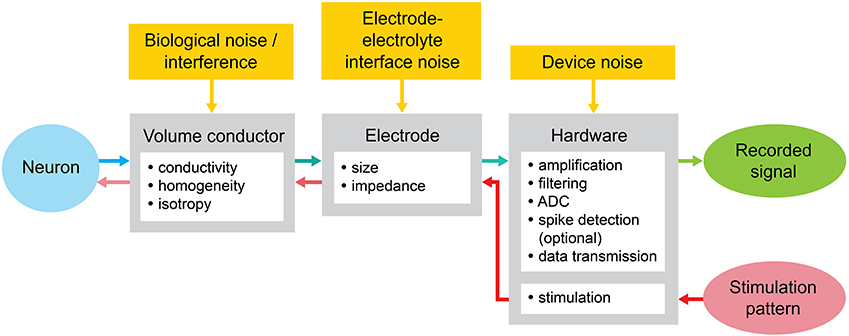
In addition to inherent noise coming from the source, i.e. the neuron, as previously described, there are several other sources of additive noise in an extracellular recording coming from the biological interface, the electrode interface and the device employed for recording. The biological noise contains unwanted electrical signatures including action potentials, subthreshold potentials and aggregate activity from distant sources. The noise collected from the extracellular environment can thus be associated to the entire recording and not to an individual neuron due to the spatial averaging in the medium and it cannot be separated with currently available methods. The interface between the liquid extracellular medium and the rigid substrate generates noise following a characteristic frequency 1/? and 1/?` [40, 41], in addition to thermal noise proportional to the electrode impedance. Furthermore, the electronic device employed for amplification, filtering, digitization and transmission can also add noise to the recorded signal. In summary, at every step of acquisition and processing (Figure 2.1) noise can be incorporated in the recording .The recordings in MEA implants are considered ideal if an experimenter can easily identify spike waveforms. However, in addition to all the noise sources previously described, common problems during the experiment may arise due to faulty channels or channels carrying spurious signals. This problem is exacerbated in long term chronically-implanted depth electrodes, where the signals begin to present much lower SNR due to unwanted factors as time progresses (e.g. electrode’s surface damage produced by the user or inflammation in tissue surrounding electrode), thereby adding difficulty to the discernment of good channels from faulty channels. To date, most work has focused on processing techniques to reliably identify neurons and evaluate integrity of electrophysiological recordings offline. However, this is not feasible for closed-loop paradigms such as spike-triggered experimental settings, wherein following a quick visual quality control check performed by the user, the recordings are further processed and reference points for the experimental framework are selected. Despite all nuisances, multichannel electrophysiology with MEAs remains to be a fundamental technique to measure the activity of several neurons simultaneously.
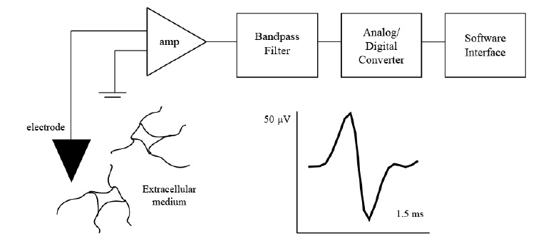
Figure 2.1. Extracellular recording experimental set-up. Extracellular recordings are collected from electrodes typically placed in the vicinity of certain neurons. The recorded signals are amplified with respect to ground, filtered, and digitized to be sent to a software interface for further processing. A typical action potential recorded
from the extracellular medium lasts 1-3 ms and has a depolarization, repolarization, and hyperpolarization period.
