Tissue Imaging With Musculoskeletal Ultrasound
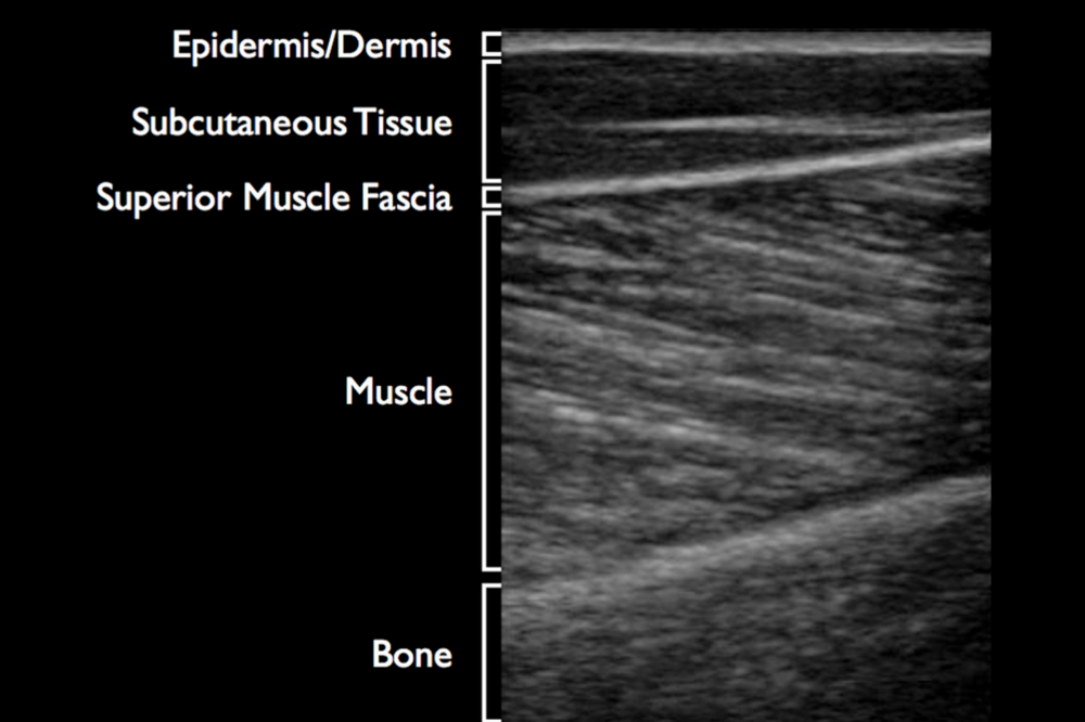
Each type of tissue in the body has a different reflectivity on an ultrasound image. This allows for differentiation between structures and helps identify landmarks. Individual tissues are characterized by the amount of reflected energy received by the machine, or known as echogenicity. More echogenic structures include bones and gas-like substances, while less echogenic structures are usually fluids. The echogenicity of tissue under MSK US imaging decreases from bone to tendon to ligament to nerve, and finally to muscle The most common structures examined during MSK US are ligaments, tendons, and muscles. Ligaments connect bones to other bones, and therefore can be seen connecting joints. Ligaments appear hyperechoic and striated. Longitudinal viewing of the ligament will produce the best view; while a transverse axis may result in a hypoechoic appearance due to surrounding hyperechoic subcutaneous fat Dynamic viewing of the ligament may show gapping when force is applied to the joint. Stressing the joint can also help the examiner identify specific bands of a ligament when the ligament may have multiple parts to it For example, the UCL of the elbow has three bands, but the anterior band is the tightest when the elbow is bent. The most effective way of determining abnormalities is to compare ligaments bilaterally to check for differences.
Abnormalities can present as partial to complete tears varying on the amount of disruption in the fiber MSK US is also very commonly used to image tendons and examine abnormalities.
Tendons connect muscles to bone. The tissue can be shown as hyperechoic, with a fine fibrillar pattern in a parallel direction When viewed in a transverse plane the tendon will appear round and hyperechoic. It is important that the beam is perpendicular to the tendon. If not, the tissue can appear hypoechoic and go undiscovered during imaging. Injury to tendons can be characterized by any gapping, disruption, or thickening of the structure. A complete tear will result in an anechoic space that disrupts the parallel appearance of the tendon. Partial tears will result in a smaller anechoic space with some parallel fibers intact. A complete examination of a tendon includes both passive and dynamic scanning. Moving the patient while scanning can aid in diagnosing certain conditions that would have been overlooked. Dynamic motions can also help orient the clinician to better identify the exact location of the pathology.
The last most common type of tissue scanned by MSK US is muscle. The appearance of muscular tissue in MSK US is hypoechoic with small sections of hyperechoic tissue. The muscle is surrounded by connective tissue that appears hyperechoic and can help outline the muscle. During dynamic scanning, muscle contraction will show movement in the tissue that can assist in identifying the individual muscle being scanned. Similar to tears in tendons, muscle tears can be identified by hyperechoic or hypoechoic fluid buildup and hypoechoic disruption in the parallel pattern of the muscle depending on the level of damage. Grade one tears will present with no disruption as compared to a grade two and three strains, which will present with partial tears or complete disruption of the fibers Blunt force injuries will show pockets of hypoechoic hematoma in the tissue that can be outlined by the torn edges of the affected tissue Musculoskeletal ultrasound can also be used to identify scar tissue in the muscle that did not heal correctly. The scar tissue appears hyperechoic and unchanged during muscle contraction.
Tissue Imaging With Musculoskeletal Ultrasound
