Trigeminovascular Anatomical Pain Pathway
Although precedence of either vascular or neuronal components have not been determined from clinical and experimental studies, the trigeminal system has been implicated in the generation of migraine pain (Tajti, 2012). Pain sensitivity in the skull
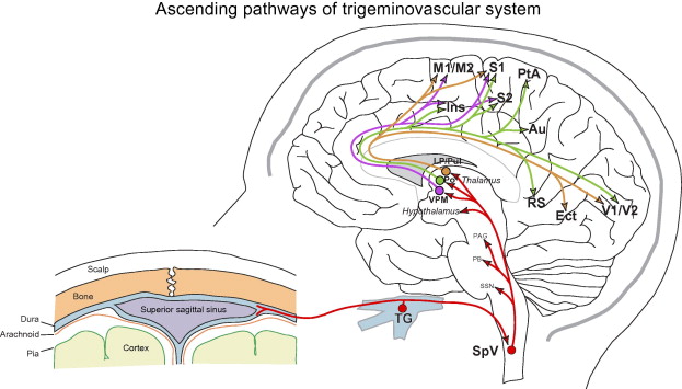
mainly occurs in large cerebral blood vessels, blood vessels in the pia and dura mater, and large venous sinuses which are surrounded by the peripheral branches of nonmyelinated (C-fibers) and thinly myelinated (Aδ fibers) axons that have vasoactive neuropeptides from the ophthalamic division of the trigeminal ganglion and in the posterior fossa from upper cervical dorsal roots (Akerman, 2011; Pietroban & Striessnig, 2003; Tajti, 2011). CSD, CGRP (calcitonin-gene related peptide), PACAP (pituitary adenylate cyclase activating peptide), and NO (nitric oxide) activate the peripheral branches of the trigeminal nerve. The afferents from the trigeminal ganglion and from the greater occipital nerve through the cervical ganglion project to second-order neurons in the brainstem in an area called trigeminal cervical complex (TCC) (Akerman, 2011; Tajti, 2011). There is a convergence of intracranial and extracranial primary afferents onto the TCC that may contribute to pain perception specified in the periorbital and occipital regions (Noseda & Burstein, 2013). The TCC neurons travel through the uintothalamic ract (carries sensory information from the head and face through the trigeminal nucleus), cross in the brainstem, and synapse with the thalamus. Functionally identified TCC neurons project to the anterior hypothalamus, lateral hypothalamus, perifornical hypothalamic areas, parabrachial area, zona incerta, lateral preoptic nucleus, and ventral posteromedial (VPM), posterior (Po), and parafascicular (Pf) thalamic nuclei. Most of the second-order TCC projections end in the ventrolateral periaqueductal gray matter (PAG), rostral trigeminal spinal nuclei, nucleus of solitary tract, brainstem reticular areas, and the cuneiform nuclei (Noseda & Burstein, 2013). Also, parasympathetic outflow to the cranium occurs from the TCC neurons connected to the superior salivatory nucleus (SSN) in the pons which links to cranial vasculature through the sphenopalatine ganglion (Akerman, 2011). TCC projections to brainstem structures (i.e. LC and PAG) and higher order structures (i.e. thalamus and hypothalamus) then send signals to the cortex. The thalamus is the main area where nociceptive inputs go through to reach the cortex. VPM durasensitive neurons travel to the primary and secondary somatosensory cortices and the insula which indicates involvement in sensory-discriminative migraine elements like intensity, location, and quality of pain. Other dura-sensitive projections from the posterior (Po) and lateral posterior/dorsal thalamic nuclei end in several cortical areas like somatosensory, visual, auditory, motor, parietal, retrosplenial, and olfactory cortices. These areas show involvement with allodynia, photophobia, phonophobia, motor clumsiness, difficulty focusing, transient amnesia, and osmophobia. The cortex sends descending projections to the thalamus, hypothalamus, and LC. Hypothalamic nuclei, such as the A11 dopaminergic nucleus, project to the TCC neurons. Also, the TCC receives descending modulation from the PAG through the rostral ventromedial medulla (Akerman, 2011).
Trigeminovascular Anatomical Pain Pathway
