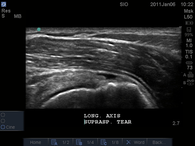
Musculoskeletal Ultrasound
Musculoskeletal ultrasound (MSK US) has been used to diagnose medical conditions for many years, but only recently has been used by sports medicine clinicians Advances in technology have increased the quality of images produced to the level of modern magnetic resonance imaging (MRI) machines.7 The modality uses sound waves transmitted through a probe to produce an image. Typically, frequencies from 5 to 20 million cycles per second (MHZ) are used Higher frequencies create better resolution, but also reduce the depth of the imaging field To create the image, pulses of ultrasound from the transducer produce echoes at tissue or organ boundaries. The waves are absorbed and reflected by each individual type of tissue differently, causing differences in appearance in the image The differences in appearance can be categorized by the term echogenicity. The echogenicity, or capacity of a structure to reflect back sound waves, can be categorized into three groups, hyperechoic, hypoechoic, and anechoic.
Hyperechoic tissue shows a high reflective pattern and appears brighter than surrounding tissue. Hypoechoic tissue has a lower reflective pattern, and shows areas with tissue that is not as bright as surrounding tissue. Finally, anechoic tissue does not show any reflectivity and therefore appears black in the image Acquiring images requires exposed skin and a medium, usually a water based gel, to facilitate surface adherence.
Benefits of Musculoskeletal Ultrasound
The use of MSK US has become more common as a primary diagnostic tool. Images that are acquired through MSK US are comparable in quality to MRI, which has been the gold standard in imaging for the musculoskeletal system Unlike an MRI, MSK US has the ability to capture real-time, dynamic images while the patient is moving. This allows clinicians to observe tissues at different lengths and in different positions, and may provide a more comprehensive exam of the injured body part. Dynamic images also give physicians an opportunity to perform guided injections and assist in fluid collection to ensure correct placement of the needle Compared to other imaging techniques, the use of sound waves does not prevent individuals with pacemakers, cochlear implants, or magnetic artifacts in the tissue from receiving the scans, and does not have any known contraindications for treatment MSK US machines are also much cheaper than other imaging modalities such as MRI’s and CT scan, and can be portable which helps decrease insurance costs to patients.
Technique and Limitations
MSK US can be ineffective at diagnosing pathologies if the operator is poorly trained in the use of the machine. Understanding the technology is important, but technique when imaging is the most important factor in getting high quality images to provide an accurate diagnosis Nofsinger et al developed an algorithm for clinicians to use before obtaining images to provide uniformity during all scans. The steps involve establishing the left versus the right end of the probe, using a bony landmark as a reference point when scanning, and stabilizing the angle of the probe once on the skin to maintain a steady orientation. The clinician should constantly be aware of their body position and angle of the probe to provide the most accurate picture possible
on the machine. A slight change in angle or pressure can completely reverse the appearance of certain tissue. This is the concept of anisotropy, which can cause the clinician to perceive incorrect findings due to a change in echogenicity. To prevent this, a firm grip on the transducer is recommended, while also placing two or three fingers on the skin to increase contact on the skin to detect minor changes in angle Another factor that can affect the appearance of tissue is the patient’s body position. Correct posture is important, especially when examining the shoulder. Forward rounding of the shoulders will cause increased difficulty in finding the structures the clinician is examining. With this in mind, proper patient position is an important factor in ultrasound technique because it will provide the clinician with the best possible image. Diagnosis is dependent on the operator being able to find the area of injury and also identify the extent of damage to the tissue.8,9 If the operator is not experienced, they may overlook defects that would be crucial to a diagnosis. Also, as technology evolves, the operator must as well. Continuing education is a very important factor in providing the best possible care.
Another limitation is the amount of penetration achieved by the acoustic waves. Deeper structures are much more difficult to examine because of weaker signals received by the machine. The more tissue the waves pass through, the less clear the return image is. This is why MSK US is rarely used to diagnose hip and spinal pathologies. Bone reflects all sound waves and will most likely block out surrounding structures due to decreased transmission of the waves. To perform an accurate examination with MSK US, the examiner must have proper training on the machine and also understand limitations of the modality.

Musculoskeletal Ultrasound

