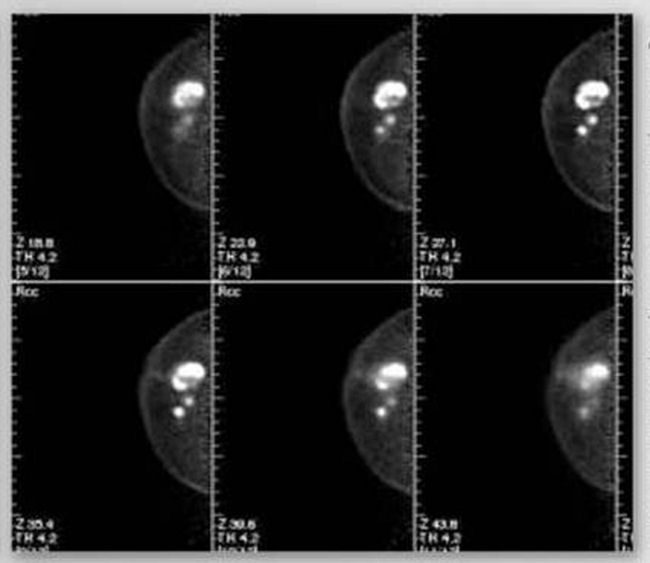
Micro-CT in Breast Cancer and Margin Assessment
Micro-CT scans of lumpectomy specimens are another proposed solution to intraoperative margin assessment. Micro-CT is a technology which allows for 3-dimensional imaging of smaller specimens, providing faster imaging than traditional CT scans. Resolution of the scan is most often inversely related to specimen size. The 3D images can be visualized as volumes that can be rotated and translated, or as 2D “slices” in any orientation. This circumvents the orientation issue posed by specimen radiographs. Tang et al. (2013) provided one of the first looks at intraoperative Micro-CT scans of lumpectomy specimens. Using a 15 minute scan/reconstruction protocol, they were able to scan 46 lumpectomy specimens, and successfully identified 32 of 55 positive margins (60% sensitivity, 93% specificity). All determinations of benign and malignant tissue were interpreted based on structure and shape.
Micro-CT in Breast Cancer and Margin Assessment
Gulfer et al. (2011) looked at micro-CT scans of breast core needle biopsies, which are very small specimens that allow for high resolution scans. The samples, amongst which the largest single dimension measured 1.3cm, were scanned in a paraffin cassette with a voxel size of 8 microns for up to 36 minutes. Samples were then fixed in paraffin, serial-sectioned, and roughly registered against their corresponding micro-CT images. Regions of interest in the micro-CT images, verified by histopathology and including fat, fibrosis, and tumor, were sampled and compared to detect differences in x-ray attenuation (i.e differences in radiodensity). For the given scan time and resolution, every tissue comparison had a statistically different radiodensity (95% confidence interval), with the exception of benign fibrosis and healthy parenchyma.
