Computer-aided diagnosis (CAD)
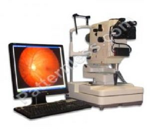
Figure 1.1: An Ophthalmic imaging system for the Retina (anterior portion of the eye). The acquired image is called a fundus image.
has become a vital part of medical evaluations and pathology detection in the United States [1]. Systematic use of CAD systems since 1980s
has caused a signicant change in the utilization of the computer output for pathology diagnosis, disease prognosis and treatment prioritization. Automated pathology detection and screening systems assist in interpretation of the medical signals, achieving a baseline evaluation and automatically analyzing disease severity, which in turn helps to
prioritize patients for treatment follow-ups. Examples of popular CAD systems include analysis of X-ray, Magnetic Resonance Imaging (MRI) and Ultrasound images.In this work, automated algorithms are presented for the analysis of two separate modalities of ophthalmic images. These images capture the human retina, or the ante-
rior part of the eye that is sensitive to light and that triggers nerve impulses of vision that are carried to the brain by the optic nerve. Retinal pathologies may be triggered as symptoms of prolonged diseases like diabetes, hypertension, high cholesterol etc.,
causing blood vessel leakage or hemorrhaging, impacting the structure and functional ities of the retinal arteries or veins, accumulation of lipids on the retinal surfaces or impacting the axiomatic plasma. If untreated or undiagnosed in a timely manner, such retinal abnormalities can lead to acquired blindness. Examples of change in vision due to retinal pathologies are shown in Fig. 1.2. The rst modality of retinal images analyzed in this work is retinal fundus imaging. Fundus images typically include the optic disc (OD, head of the optic nerve), macula (the spot which is most sensitive to vision) and blood vessels as shown in Fig.1.3

Figure 1.2: Changes in vision due to retinal pathologies
[Source:http://www.ucdenver.edu].

Figure 1.3: Example of fundus images. (a) Anatomy of a fundus image. (b) Abnormalities in fundus image due to DR.
bright and red abnormalities that occur as manifestation of non-proliferative diabetic retinopathy (NPDR) using automated algorithms can help screening and prioritization of patients based on disease severity.