Capillary phantom construction
To test whether separate strands of fibers within a large parallel bundle can be distinguished from each other, a capillary fiber phantom was created using flexible fusedsilica capillary tubing manufactured by Polymicro Inc. (Molex, Phoenix, AZ). The fibers used had a 20um inner diameter and a 90um outer diameter (including the 12um flexible polyimide coating). These fibers have been previously successfully used by Tournier et
- to construct a phantom for validating spherical deconvolution methods against q-ball
imaging. An obvious advantage of using such a fiber phantom was that it could be created to pre-defined specifications having a fully known structure. The fibers were manually cleaved and bundled with strands of human hair to have an approximate diameter of 0.6mm with about 0.1mm gap between individual bundles. The result was a large fiber bundle with approximately 2mm diameter, consisting of 14 individual fiber strands, each consisting of 48 capillary tubes with a length of 40mm. This bundle was mounted on a white high density polyethylene (HDPE) carrier and affixed using dental floss. The carrier was then wedged into a specimen tube made of transparent polyethylene (PE) plastic with 10mm diameter and 45mm length. A photograph of the phantom before immersion in water is shown in figure1. The left panel (a) shows the bundle mounted on the carrier, the right panel (b) shows the carrier inserted into the specimen tube. The tube was then filled with medium (medium recipe detailed below) and put upright on a shaker to be vibrated for several hours, making sure all the air would be purged from the capillary tubes. This process was tested beforehand on a single fiber and validated on a light microscope to confirm that the capillaries can be filled with water. The cap was then closed submerged in a beaker of medium, making sure to purge all the air when closing it. The tightly closed specimen tube was suspended with fishing line in a white PE plastic tube with approximately 100mm diameter and 25mm length (MR localizer showing structure in figure 2). The tube was filled with medium and air purged. The medium was made according specifications for the fBIRN agar phantom following the protocol by Schneiders but leaving out the agar (which would not be taken up the capillary tubes).
Medium (3L):
- 2700ml H2O (deionized)
- 300ml 21.8mM NiCl2 stock (to adjust T1/T2 relaxation time)
- 15g NaCl (for conductivity)
- 0.75g NaA (to inhibit bacterial growth)
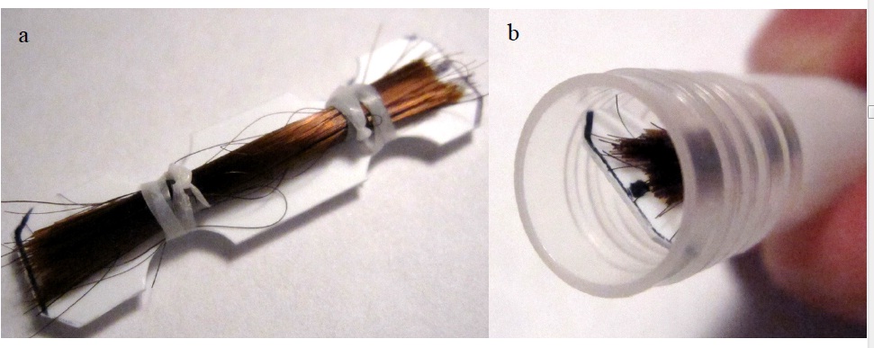
Figure 1 Pictures of the capillary fiber phantom: Fiber bundle mounted on the carrier
(panel a, left) and inserted into specimen tube (panel b, right).
Figure2 MRI localizer images of assembled phantom: The specimen tube with the fiber
bundle is suspended in the outer PE plastic tube using fishing line. Imaging orientations are axial (panel a), coronal (panel b), and sagittal (panel c). The red arrow points to thespecimen tube inside the PE tube.