Bone microarchitecture and structure in patients with AS
Previous studies on bone microarchitecture and structure in patients with AS
Not much is known about the microarchitecture of bone in AS. In 1992, Devogelaer and colleagues studied 10 patients with AS by using quantitative computed tomography (QCT) to image the lumbar spine. The results revealed that AS patients had low trabecular bone density in the vertebral bodies (Devogelaer 1992). The effect was more marked in those with severe AS, syndesmophytes and apophyseal joint fusion. In another small study (n=15) done using spine QCT, a statistically significant decline in trabecular bone mineral content (BMC) was observed (change ± SD: 18 ± 7 mg/cm3) over a period of ten years (Korkosz 2011). Patients with advanced spine involvement had significantly lower BMC both at baseline and follow-up (Korkosz 2011). However CT based measurements of the spine has certain limitations. First, CT based measurement is processed from the middle layer of the vertebra and hence may not give information about the structure of whole vertebral body. Second, the error rate because of interference from fat is high (SE: 10-30%). Third, it is also not possible to analyze a fractured vertebra. Finally, the radiation exposure is also higher (SE-QCT <100 IS).
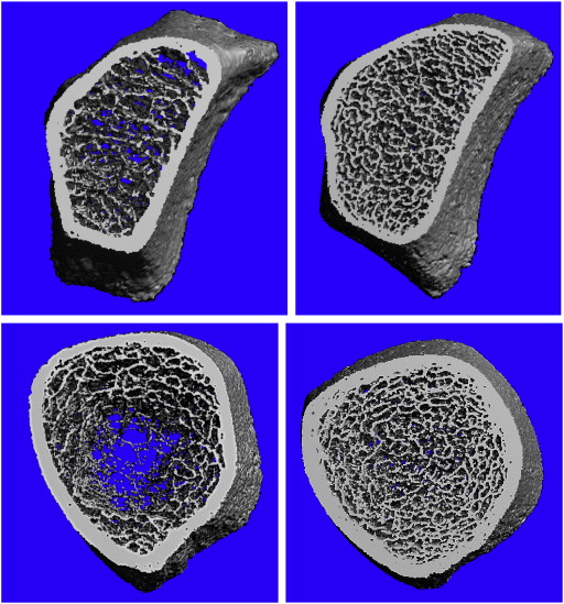
Abnormal bone microarchitecture, strength and biomechanics in rat models of spondylitis
In 2009, Rauner and colleagues studied bone quality in the tibia and sixth vertebral body of HLA B27 rats and demonstrated abnormal trabecular structure in AS (Rauner 2009). The diseased rats had significantly reduced trabecular thickness and number, and increased trabecular separation. Similar results were observed in another study on transgenic rat models (Dempster 2001).
Bone microarchitecture and structure in patients with AS
