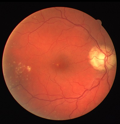
All classification systems perform image processing to extract features that we hope will make it easier for the software to correctly label each pixel as vessel or not vessel, however unsupervised systems (like this work) do not require additional training data to develop the classification criteria to assign the label. Sometimes this is simply because the decision criteria is implicit or hand-tuned (e.g., a hand chosen threshold value).
One of the earliest approaches published used matched filters at 12 orientations to approximate linear segments of blood vessels at all orientations [4] unlike the other papers, this was implemented in hardware and takes a very classical signal processing approach to extracting vessels, its strengths are its simplicity. The method (along with hand-tuned parameters) builds a relatively simple model that still produces qualitatively good results. It does fail to account for illumination conditions both uneven illumination, and the central vessel reflex.
Zana and Klein applied sophisticated morphological processing based on a Gaussian model of (the profile of) blood vessels these included a sum of directional top-hat filters and what they call geodesic reconstruction [25] this amounts to a sum of top hat filters then filtering noisy segments by curvature (using the Laplacian). [7] dealt with the uneven illumination and irregular vessel structures using an adaptive thresholding scheme with a verification procedure to reject non-vessel points.
The verification procedure is where the prior information about the shape characteristics of the blood vessels are introduced. Specifically, for each chosen threshold they calculate the distance transform they then “prune” the result to find the vessel centerlines using critera based on angle, width, contrast and a minimum size. They then reconstruct the final image by taking the logical OR of all of the processed images at each threshold value. Finally they post-process the images using non-maximal suppression by calculating the gradient magnitude and orientation using the Sobel operator and then for each orientation removing the pixels that do not have the greatest gradient magnitude.
[11] implemented another variant on morphological reconstruction. They preprocess the image to account for irregular illumination by subtracting the resulting image from convolving with a large arithmetic mean kernel, then enhance small vessels by adding the strongest result of convolving the image with line detecting features at 0, 45, 90, and 135 degrees at each pixel. Like [7] they then extract center lines, in this case with 4 Difference of Offset Gaussian filters which are then connected by region growing and filtered by a validation procedure. The authors then construct a multiscale representation using the morphological top-hat filter (albeit a modified version, to try to suppress noise) using circular structuring elements with radii from 1 to 8 pixels. Finally the authors perform another round of vessel filling using a so-called “Double Threshold operator” and post-process by considering pixels with 6 out of 8 connected neighbors that are vessel pixels to also be labeled as vessel pixels.
The unsupervised approaches are very similar to each other in that they each attack the known difficulties with these images and attempt to use the geometry of the vessels to guide the labeling of pixels. Every paper so far has applied operations to compensate for uneven illumination and/or aperture effects, find the center-line of the vessels and then, having identified the most easily labeled vessel pixels they then extend out to try to label the rest of the vessel (this is another challenge of this data set, it is not sufficient to generate thin line boundaries, but one must instead densely label the vessel).
An important disadvantage of the proposed methods is that most of them take ad-hoc approaches to trying to find the paths of the vessels, as a result they have many thresholds and parameters that must be tuned (and in general which hurt the prospects that these systems could ultimately be deployed in the real world with varying hardware and environmental conditions). Additionally we have seen that the methods above assume an explicit model (e.g., Gaussian intensity profile ) for the shape of the vessels. We hope to show in this thesis that with very simple feature extraction and the Tensor Voting framework that we have implemented a system that is conceptually similar to these related works, but which naturally and elegantly expresses these constraints on shape while being model-free with respect to the shape of the vessels (and which can easily be extended to 3D data).
[4] S. Chaudhuri, S. Chatterjee, N. Katz, M. Nelson, and M. Goldbaum, “Detection of blood vessels in retinal images using two-dimensional matched filters,” IEEE Transactions on medical imaging, vol. 8, no. 3, pp. 263–269, 1989.
[7] X. Jiang and D. Mojon, “Adaptive local thresholding by verification-based multithreshold probing with application to vessel detection in retinal images,” Pattern Analysis and Machine Intelligence, IEEE Transactions on, vol. 25, no. 1, pp. 131–137, 2003.
[11] A. M. Mendonca and A. Campilho, “Segmentation of retinal blood vessels by combining the detection of centerlines and morphological reconstruction,” Medical Imaging, IEEE Transactions on, vol. 25, no. 9, pp. 1200–1213, 2006.
[25] F. Zana and J.-C. Klein, “Segmentation of vessel-like patterns using mathematical morphology and curvature evaluation,” Image Processing, IEEE Transactions on, vol. 10, no. 7, pp. 1010–1019, 2001.


