Computed Tomography (CT)

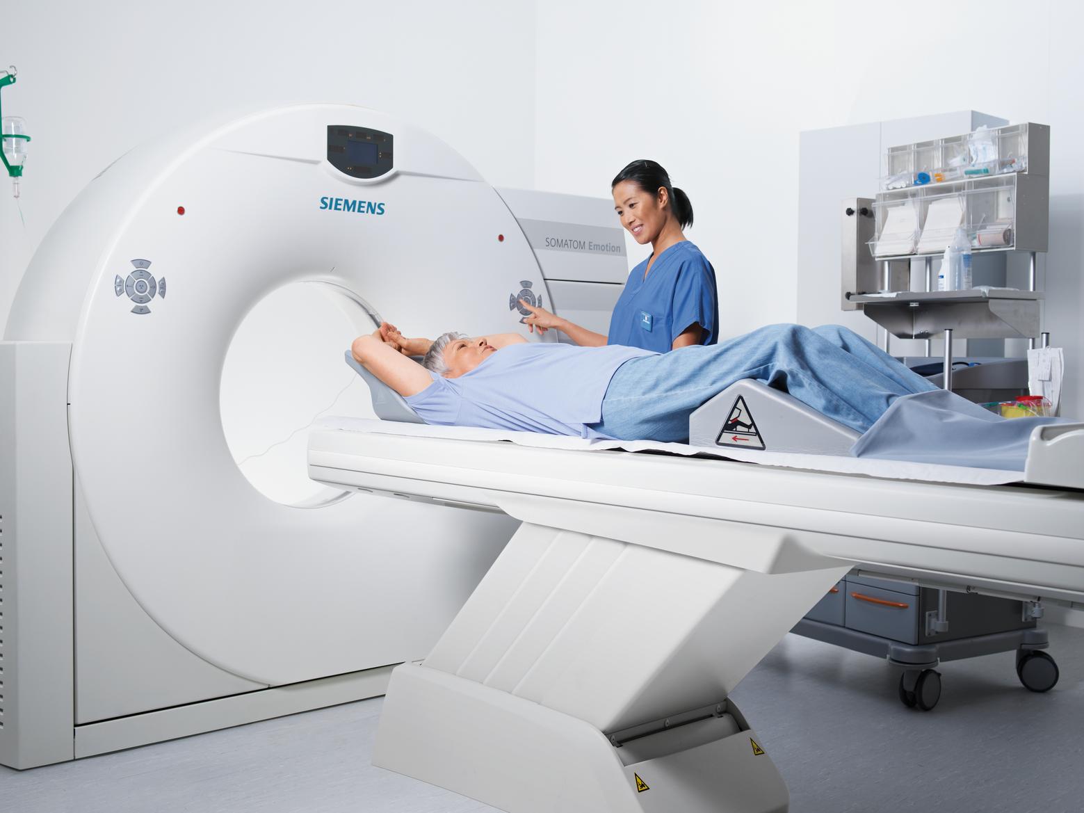
X-ray Computed Tomography (CT) is a medical imaging technique that enables the reconstruction of cross-sections of an object, using a series of X-ray measurements taken from many angles around the object. The first CT scanner was introduced by Sir Godfrey Hounsfield (1972) at EMI (London, England), for which, Alan Cormack and Hounsfield were awarded the Nobel Prize at 1979 . Since then, CT scanners have been improved both mechanically and electronically enabling high resolution CT images to be generated and displayed very quickly. The current quality and speed with which they are generated is in part due to improvements in efficient image reconstruction algorithms. Although the problem of tomographic image reconstruction was mathematically solved by Johann Radon in 1917 the first scanner used a simple algebraic reconstruction algorithm, since the connection between the new modality and the Radon transform was unknown at that time. This field is still evolving and new algorithms are currently used that adapt to the new projection geometries and new mathematical theories.In addition, iterative reconstruction algorithms, which enable major radiation dose reductions,have been developed. A variety of CT projection geometries, shown in figure 2 and the classical
single step and iterative CT reconstruction algorithms are briefly discussed in this chapter

Figure 2: Three X-ray source geometries: (A) Parallel Geometry, (B) Fan beam Geometry,
and (C) Cone beam Geometry.
For more than a century conventional X-ray radiography has offered a valuable non-invasive means of diagnosis, but, it suffers from a number of limitations. For instance, imaging the brain with radiography typically yields insufficient details. This is due to the fact that each picture element displays the sum of all attenuation components along the ray lines. As a result, the contrast of the images are dominated by the structures with high attenuation, such as bone, and thereby, the low intensity objects, such as soft tissues, are completely hidden in most cases.This issue is solved through the development of cross-sectional imaging modalities, such as Computed Tomography (CT), in which the neighboring and superimposed structures have very little influence on the image.Over the past two decades, there has been a major increase in the clinical use of CT primarily due to its unsurpassed speed and the fine detail that can be obtained in cross-sectional views of the internal organs. However, compared to conventional radiography, CT results in a relatively large radiation dose to patients. Studies over the past decade have shown that the higher radiation dose is of serious long-term concern for increasing the potential risk of developing cancer. Based on comparable risk estimates along with information on CT scan usage from 1991 through 1996, about 0.4% (or 4 in 1000) of all cancers in the United States may be related to radiation from CT scans. For older people, this issue is not very serious, since cancers that could result from CT scan-related radiation exposure would take years to develop. However, given the fact that there is increased use of CT scans in the pediatric population, this could be a problem since these children can be expected to be alive many years after a scan was performed. As a result, low dose CT imaging that maintains the resolution and achieves good contrast to noise ratio has been the goal of many CT developments over the past decade.
CT image quality
CT image quality is usually described in the sense of contrast, spatial resolution, image noise, and artifacts. Different metrics have been used to measure the performance of the physical systems and the reconstruction algorithms. Signal to noise ratio (SNR) and contrast to noise ratio (CNR) are usually used as surrogate measures for CT image quality. Other metrics are also used to measure the effect of detectors, reconstruction and post-processing algorithms on the images, such as modulation transfer function, noise power spectrum, noise equivalent quanta, and detector quantum efficiency. More accurate metrics have been recently used to model the observer performance with a good compatibility with the human observer studies, such as model observer methods and detectability index
One of the most important applications of CT

One of the most important applications of CT is in detecting low contrast structures, a task that is limited primarily by image noise and therefore by the radiation dose: a higher radiation dose results in a lower noise, thereby improving the contrast detectability. In one of the simplest definitions, the CT image noise is the standard deviation of voxel values in a homogeneous (typically water) phantom, and this is influenced by a large number of parameters, including:
1. Electronic noise: caused by electronic circuits and the X-ray detectors.
2. Statistical noise: resulting from fluctuations in the the number of X-ray photons reaching the detectors and detected by them. This can be controlled by :
Radiation dose, which itself is a function of X-ray source peak voltage (kVp), the product of X-ray source current and exposure time (mAs), table speed in helical CT exams (pitch), X-ray collimation and exposure area. Reconstruction slice thickness, changing the thickness changes the number of detected X-ray photons. For example, compared with a slice thickness of 5 mm, a thickness of 10 mm approximately doubles the number of X-ray photons entering each detector.
3. The choice of reconstruction and filtering algorithms.
4. Artifactual noise: the presence of artifacts might be viewed as a form of noise that interfere with the interpretation of the CT image, for instance beam hardening, streak artifacts and photon starvation effects.

